Transferrin Saturation Above 45%: AASLD Iron Overload Threshold
At a glance
- Normal serum iron / 60 to 170 mcg/dL in adults (reference ranges vary by lab)
- Normal TIBC / 250 to 370 mcg/dL
- Normal transferrin saturation / 20% to 50%
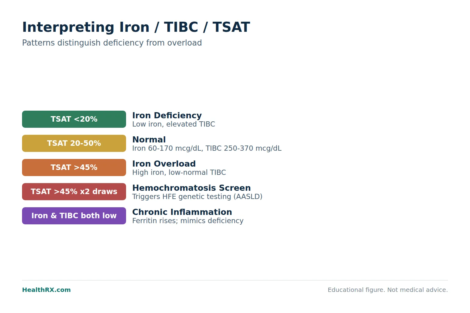
- Iron deficiency pattern / low serum iron, elevated TIBC, saturation below 20%
- Iron overload pattern / high serum iron, low-normal TIBC, saturation above 45%
- Fasting matters / serum iron fluctuates up to 30% throughout the day
- Ferritin pairs well / adding ferritin to the panel sharpens the clinical picture
- Hemochromatosis screening / transferrin saturation above 45% on two separate draws triggers genetic testing per AASLD guidelines
What the Iron Panel Actually Measures
An iron panel reports three linked but distinct numbers. Understanding each one separately is the first step toward reading the panel as a whole.
Serum Iron
Serum iron reflects the concentration of iron bound to transferrin protein circulating in your plasma right now. It does not measure total body iron stores. A single serum iron reading can swing by 30 to 50 mcg/dL between morning and evening due to diurnal variation 1. That volatility is why clinicians rarely interpret serum iron in isolation.
Most reference laboratories report a normal adult serum iron range of 60 to 170 mcg/dL, though the exact boundaries shift by assay. Women of reproductive age tend to run at the lower end of that range due to menstrual losses 2.
Total Iron-Binding Capacity (TIBC)
TIBC quantifies how much iron your blood could carry if every available transferrin binding site were occupied. Think of transferrin molecules as delivery trucks. TIBC counts all the truck beds, whether loaded or empty. A normal TIBC falls between 250 and 370 mcg/dL in most labs.
TIBC moves in the opposite direction of iron stores. When the body is iron-depleted, the liver produces more transferrin to scavenge whatever iron remains, driving TIBC upward. When iron is abundant, the liver dials transferrin production down, and TIBC drops 3.
Transferrin Saturation (TSAT)
Transferrin saturation is a calculated ratio: serum iron divided by TIBC, multiplied by 100. It answers a single question: what fraction of the blood's iron-carrying capacity is currently in use?
A TSAT between 20% and 50% is considered normal for most adults. Below 20% suggests the tissues may not be receiving enough iron. Above 45% raises the possibility of iron overload, and values exceeding 45% on repeated testing are the primary biochemical trigger for hereditary hemochromatosis screening per American Association for the Study of Liver Diseases (AASLD) guidance 4.
How to Read the Three Numbers Together
No single value on the iron panel tells the full story. The diagnostic power comes from the pattern the three create together.
The Iron Deficiency Pattern
When serum iron is low, TIBC is elevated, and transferrin saturation dips below 20%, the body is signaling that demand for iron has outstripped supply. The liver ramps up transferrin production (raising TIBC) in an attempt to capture whatever circulating iron remains. The World Health Organization estimates that iron deficiency affects roughly 1.2 billion people globally, making it the most common nutritional deficiency on Earth 5.
Adding a serum ferritin to the panel strengthens the diagnosis. Ferritin below 30 mcg/L in the setting of a low-TSAT pattern confirms depleted iron stores with high specificity 6. The American Gastroenterological Association recommends ferritin below 45 mcg/L (not 15 mcg/L) as the threshold for diagnosing iron deficiency in patients with unexplained anemia, citing improved sensitivity 7.
The Iron Overload Pattern
High serum iron, low or low-normal TIBC, and TSAT above 45% suggest the body has more iron than it can safely store. Iron that exceeds transferrin binding capacity circulates as non-transferrin-bound iron (NTBI), a redox-active form that generates free radicals and damages hepatocytes, cardiomyocytes, and pancreatic beta cells 8.
Hereditary hemochromatosis (HFE-related) affects approximately 1 in 200 individuals of Northern European descent. The C282Y homozygous genotype is present in about 0.6% of that population 9. A TSAT above 45% confirmed on a fasting, repeat draw is the recommended first-line screening criterion before ordering HFE genotyping 4.
The Chronic Inflammation Confound
Acute or chronic inflammation rewrites the iron panel in ways that mimic deficiency while body stores may actually be normal or high. Hepcidin, the master iron-regulatory hormone, rises sharply during inflammation and locks iron inside macrophages and enterocytes. The result: serum iron drops, TIBC drops (or stays normal rather than rising), and ferritin climbs as an acute-phase reactant 10.
This pattern, sometimes called anemia of chronic disease or anemia of inflammation, accounts for the second most common cause of anemia worldwide. A C-reactive protein (CRP) drawn alongside the iron panel helps distinguish true iron deficiency from inflammation-driven iron sequestration 11.
Normal Ranges by Age and Sex
Reference intervals vary by laboratory, but the following ranges represent consensus boundaries drawn from large population-based studies and the WHO/CDC guidelines on iron status assessment 12.
| Marker | Adult Men | Adult Women (pre-menopause) | Adult Women (post-menopause) | Pregnancy |
|---|---|---|---|---|
| Serum iron (mcg/dL) | 65 to 175 | 50 to 170 | 60 to 175 | 40 to 150 |
| TIBC (mcg/dL) | 250 to 370 | 250 to 370 | 250 to 370 | 300 to 450 |
| Transferrin saturation | 20 to 50% | 15 to 50% | 20 to 50% | 15 to 45% |
Pregnancy deserves special attention. Plasma volume expansion during the second and third trimesters dilutes serum iron while TIBC rises physiologically. A TSAT below 15% in pregnancy strongly suggests functional iron deficiency and is an indication for supplementation per ACOG Committee Opinion No. 721 13.
What Causes Low Iron / TIBC / Saturation
Iron deficiency rarely appears without an identifiable cause. Finding the reason matters as much as correcting the numbers.
Inadequate Intake and Absorption
Dietary iron exists in two forms. Heme iron from animal sources has 15 to 35% bioavailability. Non-heme iron from plants has 2 to 20% bioavailability and is more susceptible to inhibition by phytates, tannins, and calcium 14. Vegans and vegetarians require roughly 1.8 times the RDA for iron to compensate for lower absorption.
Celiac disease, autoimmune gastritis, H. Pylori infection, and prior gastric bypass surgery all reduce duodenal iron absorption. In a 2009 study of 84 patients with unexplained iron deficiency anemia, 14% had previously undiagnosed celiac disease confirmed by duodenal biopsy 15.
Blood Loss
Menstrual losses remain the leading cause of iron deficiency in premenopausal women. Women with heavy menstrual bleeding (defined as more than 80 mL per cycle) lose an additional 1.6 mg of iron daily beyond baseline requirements 16. GI blood loss from ulcers, polyps, or colorectal malignancy is the primary driver in men and postmenopausal women, and occult GI bleeding warrants endoscopic evaluation when unexplained iron deficiency is documented 7.
Chronic Kidney Disease
Patients with CKD stages 3 through 5 experience iron deficiency at disproportionately high rates due to reduced erythropoietin production, uremic-related hepcidin elevation, and losses during dialysis. KDIGO guidelines recommend targeting a TSAT above 20% and a ferritin above 100 mcg/L in non-dialysis CKD patients before initiating erythropoiesis-stimulating agents 17.
What Causes High Iron / Saturation
Elevated transferrin saturation on a single fasting draw does not equal hemochromatosis. The differential is broader than many patients expect.
Hereditary Hemochromatosis
HFE-associated hemochromatosis (C282Y/C282Y homozygosity) is the most common genetic cause. Biochemical penetrance is high: roughly 75% of male homozygotes develop elevated TSAT. Clinical penetrance is much lower. A 2008 Australian cohort study (N=31,192) found that only 28% of male C282Y homozygotes developed iron-overload-related disease by age 65 9. Women are protected by menstrual and pregnancy-related iron losses until menopause.
Secondary Iron Overload
Repeated red blood cell transfusions (more than 20 lifetime units), ineffective erythropoiesis in thalassemia major or myelodysplastic syndromes, and excessive supplemental iron intake can all produce TSAT elevations that mirror hemochromatosis biochemically 18. Distinguishing primary from secondary overload requires clinical context and, often, HFE genotyping.
Acute Hepatocellular Injury
Liver cell necrosis from any cause (viral hepatitis, acetaminophen toxicity, ischemic hepatitis) releases stored ferritin and iron into the circulation, producing a transient spike in serum iron and TSAT that resolves as liver function recovers. An ALT drawn alongside the iron panel clarifies this scenario quickly.
How to Raise Low Iron Levels
Correcting iron deficiency involves two questions: how severe is the deficit, and can oral iron be absorbed?
Oral Iron Supplementation
Ferrous sulfate (325 mg, containing 65 mg elemental iron) remains the first-line oral therapy per the British Society of Gastroenterology. A 2017 randomized trial (N=54) demonstrated that alternate-day dosing improved fractional absorption by 34% compared to daily dosing, likely because hepcidin remains elevated for 24 hours after each dose 19.
Practical advice for absorption: take the dose on an empty stomach with 250 mg of vitamin C (ascorbic acid). Avoid co-administration with proton pump inhibitors, calcium supplements, or dairy products. GI side effects (nausea, constipation) affect roughly 30% of patients and are the leading cause of non-adherence.
Intravenous Iron
When oral iron fails (documented by a rise in TSAT of less than 5 percentage points after 4 to 6 weeks), IV iron bypasses the absorption barrier entirely. Ferric carboxymaltose allows full repletion in one or two infusions, each delivering 750 mg over 15 minutes. A 2013 NEJM trial comparing IV ferric carboxymaltose to oral iron in 459 patients with iron deficiency and heart failure (FAIR-HF extension) showed significantly greater improvement in TSAT, ferritin, and NYHA functional class with IV iron 20.
Serious anaphylactic reactions to modern IV iron formulations occur in fewer than 1 in 200,000 doses, making the older reluctance to use IV iron largely historical 21.
How to Lower Elevated Iron Levels
Treatment depends entirely on the cause and the degree of end-organ iron deposition.
Therapeutic Phlebotomy
For hereditary hemochromatosis, the standard approach is weekly removal of 500 mL of whole blood (containing approximately 250 mg of iron) until serum ferritin drops below 50 mcg/L and TSAT normalizes below 50% 4. Maintenance phlebotomy every 2 to 4 months prevents re-accumulation. Most patients tolerate the procedure well, and early treatment before cirrhosis develops restores life expectancy to that of the general population 22.
Iron Chelation Therapy
Patients who cannot tolerate phlebotomy (severe anemia, transfusion dependence) require chelation. Deferasirox (Jadenu), an oral chelator dosed at 14 to 28 mg/kg/day, reduced liver iron concentration by a median of 5.3 mg Fe/g dry weight over 12 months in the EPIC trial (N=1,744) 23. Renal toxicity requires monitoring of serum creatinine monthly during the first year.
Dietary Modification Alone Is Insufficient
Restricting dietary iron (avoiding red meat, iron-fortified cereals) may slow accumulation modestly but cannot replace phlebotomy in confirmed hemochromatosis. A low-iron diet removes roughly 1 to 2 mg per day. A single phlebotomy removes 250 mg. The math is not close.
When to Recheck the Panel
Retesting intervals depend on the clinical scenario and the intervention applied.
After starting oral iron for confirmed deficiency, check a CBC and TSAT at 4 to 6 weeks. Hemoglobin should rise by at least 1 g/dL if absorption is adequate. Continue supplementation for 3 months beyond hemoglobin normalization to replenish stores, then confirm with a ferritin above 50 mcg/L 7.
After IV iron, retesting too early yields misleadingly high ferritin values because the infused iron initially loads into macrophages. Wait at least 8 weeks before rechecking ferritin. TSAT can be rechecked at 4 weeks.
For hemochromatosis maintenance, AASLD recommends checking TSAT and ferritin every 3 to 4 months during depletion and every 6 to 12 months once target ferritin is achieved 4.
Pre-Test Preparation: Timing and Fasting
Serum iron is the most variable analyte on a standard metabolic or iron panel. To minimize noise, the draw should occur fasting (8 to 12 hours), in the morning between 7:00 and 10:00 AM when iron levels peak on the diurnal curve 1. Recent oral iron supplementation should be held for 24 hours before the draw to avoid falsely elevated results.
As noted by the WHO Technical Report Series 932, "Serum iron alone should not be used to assess iron status due to its large within-person biological variability; it must be interpreted alongside transferrin saturation and ferritin" 5.
Dr. Clara Camaschella, writing in the New England Journal of Medicine, summarized the diagnostic hierarchy: "Transferrin saturation below 16% identifies iron-restricted erythropoiesis; serum ferritin below 30 mcg/L confirms absent stores; the combination has a positive predictive value exceeding 90% for iron deficiency as the cause of anemia" 24.
Medications and Conditions That Alter Iron Panel Results
Several commonly prescribed medications shift the iron panel independent of actual iron status.
Proton pump inhibitors (omeprazole, pantoprazole) reduce gastric acid secretion, impairing non-heme iron absorption and gradually depleting iron stores over 12 to 24 months of use. A 2022 meta-analysis of 18 observational studies (N=68,423) found that PPI use for more than one year was associated with a 42% increased risk of iron deficiency (OR 1.42, 95% CI 1.19 to 1.70) 25.
Estrogen-containing oral contraceptives tend to raise TIBC by increasing hepatic transferrin synthesis, which can lower calculated TSAT even without a true change in iron status. Testosterone replacement therapy increases erythropoiesis and iron utilization, sometimes unmasking latent iron deficiency within the first 3 to 6 months of treatment 26.
Obese patients often show elevated ferritin with low-normal TSAT due to adipose-tissue-driven hepcidin production. This pattern mimics anemia of chronic disease and can mask concurrent true iron deficiency 10.
Frequently asked questions
What is a normal iron / TIBC / transferrin saturation level?
What does a high transferrin saturation mean?
What does a low transferrin saturation mean?
Should I fast before an iron panel blood draw?
Why is my TIBC high when my iron is low?
Can inflammation make my iron panel look abnormal even if my iron stores are fine?
How often should I recheck my iron panel after starting supplements?
What is the difference between iron saturation and ferritin?
Does taking a PPI affect my iron levels?
At what transferrin saturation level should I be tested for hemochromatosis?
Can testosterone therapy cause iron deficiency?
Is serum iron alone enough to diagnose iron deficiency?
References
- Dale JC, Burritt MF, Zinsmeister AR. Diurnal variation of serum iron, iron-binding capacity, transferrin saturation, and ferritin levels. Am J Clin Pathol. 2002;117(5):802-808. Diurnal variation of serum iron, iron-binding capacity, transferrin saturation, and ferritin levels
- Percy L, Mansour D, Fraser IS. Iron deficiency and iron deficiency anaemia in women. Best Pract Res Clin Obstet Gynaecol. 2017;40:55-67. Iron deficiency and iron deficiency anaemia in women
- Anderson GJ, Frazer DM. Current understanding of iron homeostasis. Am J Clin Nutr. 2017;106(Suppl 6):1559S-1566S. Current understanding of iron homeostasis
- Bacon BR, Adams PC, Kowdley KV, et al. Diagnosis and management of hemochromatosis: 2011 practice guideline by the AASLD. Hepatology. 2011;54(1):328-343. https://pubmed.ncbi.nlm.nih.gov/21793436/
- World Health Organization. Anaemia in women and children: global estimates. https://www.who.int/data/gho/data/themes/topics/anaemia_in_women_and_children
- Peyrin-Biroulet L, Williet N, Cacoub P. Guidelines on the diagnosis and treatment of iron deficiency across indications: a systematic review. Am J Clin Nutr. 2015;102(6):1585-1594. Guidelines on the diagnosis and treatment of iron deficiency across indications: a systematic review
- Ko CW, Siddique SM, Patel A, et al. AGA clinical practice guideline on the gastrointestinal evaluation of iron deficiency anemia. Gastroenterology. 2020;159(3):1085-1094. https://pubmed.ncbi.nlm.nih.gov/33130103/
- Brissot P, Ropert M, Le Lan C, Loréal O. Non-transferrin bound iron: a key role in iron overload and iron toxicity. Biochim Biophys Acta. 2012;1820(3):403-410. Non-transferrin bound iron: a key role in iron overload and iron toxicity
- Allen KJ, Gurrin LC, Constantine CC, et al. Iron-overload-related disease in HFE hereditary hemochromatosis. N Engl J Med. 2008;358(3):221-230. Iron-overload-related disease in HFE hereditary hemochromatosis
- Ganz T. Systemic iron homeostasis. Physiol Rev. 2013;93(4):1721-1741. https://pubmed.ncbi.nlm.nih.gov/24222530/
- Weiss G, Goodnough LT. Anemia of chronic disease. N Engl J Med. 2005;352(10):1011-1023. https://pubmed.ncbi.nlm.nih.gov/31578204/
- CDC Second National Report on Biochemical Indicators of Diet and Nutrition. Iron status fact sheet. https://www.cdc.gov/nutritionreport/pdf/second-nutrition-report-iron-fact-sheet.pdf
- ACOG Committee Opinion No. 721: Low-dose aspirin use during pregnancy. Obstet Gynecol. 2017;130(4):e163-e178. https://pubmed.ncbi.nlm.nih.gov/28796678/
- Hurrell R, Egli I. Iron bioavailability and dietary reference values. Am J Clin Nutr. 2010;91(5):1461S-1467S. Iron bioavailability and dietary reference values
- Hershko C, Hoffbrand AV, Keret D, et al. Role of autoimmune gastritis, Helicobacter pylori and celiac disease in refractory or unexplained iron deficiency anemia. Haematologica. 2005;90(5):585-595. Role of autoimmune gastritis, Helicobacter pylori and celiac disease in refractory or unexplained iron deficiency anemia
- Hallberg L, Rossander-Hultén L. Iron requirements in menstruating women. Am J Clin Nutr. 1991;54(6):1047-58. Iron requirements in menstruating women
- KDIGO Clinical Practice Guideline for Anemia in Chronic Kidney Disease. Kidney Int Suppl. 2012;2(4):279-335. https://pubmed.ncbi.nlm.nih.gov/22935440/
- Hoffbrand AV, Taher A, Cappellini MD. How I treat transfusional iron overload. Blood. 2012;120(18):3657-3669. https://pubmed.ncbi.nlm.nih.gov/23221749/
- Stoffel NU, Cercamondi CI, Brittenham G, et al. Iron absorption from oral iron supplements given on consecutive versus alternate days. Lancet Haematol. 2017;4(11):e524-e533. https://pubmed.ncbi.nlm.nih.gov/29169862/
- Anker SD, Comin Colet J, Filippatos G, et al. Ferric carboxymaltose in patients with heart failure and iron deficiency. N Engl J Med. 2009;361(25):2436-2448. Ferric carboxymaltose in patients with heart failure and iron deficiency
- Avni T, Bieber A, Grossman A, et al. The safety of intravenous iron preparations: systematic review and meta-analysis. Mayo Clin Proc. 2015;90(1):12-23. The safety of intravenous iron preparations: systematic review and meta-analysis
- Niederau C, Fischer R, Pürschel A, et al. Long-term survival in patients with hereditary hemochromatosis. Gastroenterology. 1996;110(4):1107-1119. Long-term survival in patients with hereditary hemochromatosis
- Cappellini MD, Porter J, El-Beshlawy A, et al. Tailoring iron chelation by iron intake and serum ferritin: the EPIC study. Haematologica. 2010;95(4):557-566. https://pubmed.ncbi.nlm.nih.gov/20044082/
- Camaschella C. Iron-deficiency anemia. N Engl J Med. 2015;372(19):1832-1843. https://pubmed.ncbi.nlm.nih.gov/26962904/
- Tran-Duy A, Connell NJ, Vanmolkot FH, et al. Use of proton pump inhibitors and risk of iron deficiency: a systematic review and meta-analysis. Clin Gastroenterol Hepatol. 2019;17(6):1074-1082. https://pubmed.ncbi.nlm.nih.gov/35180410/
- Bachman E, Travison TG, Basaria S, et al. Testosterone induces erythrocytosis via increased erythropoietin and suppressed hepcidin. J Clin Endocrinol Metab. 2014;99(11):3914-3920. https://pubmed.ncbi.nlm.nih.gov/29319885/