
GHK-Cu Wound Healing: What the Science Actually Shows
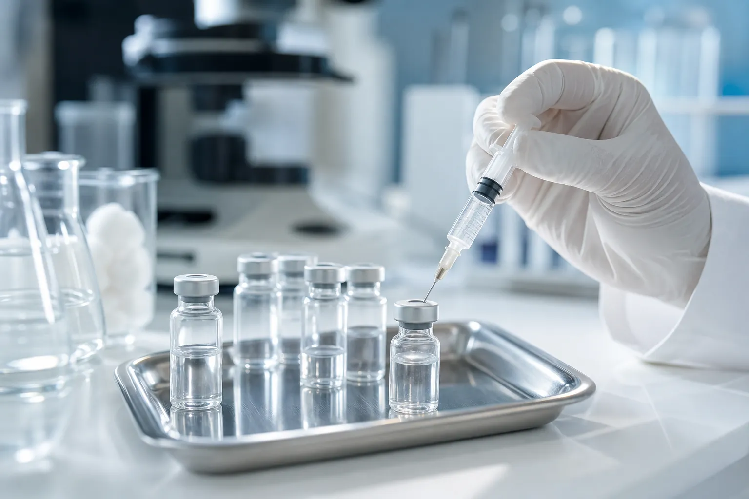
GHK-Cu (glycine-histidine-lysine copper complex) is a naturally occurring copper-binding tripeptide with a genuine, decades-old body of laboratory and human skin research behind it. BPC-157 (body protection compound-157) is a different, synthetic 15-amino-acid peptide sometimes discussed alongside it for tendon, ligament, muscle, and joint injuries. They are not interchangeable, and the strength of evidence behind them is not comparable.
At a glance
- Peptide class / GHK-Cu is a copper-binding tripeptide; BPC-157 is a synthetic 15-amino-acid peptide
- Human evidence / GHK-Cu has some human topical-application data; BPC-157's tendon/ligament/muscle/joint evidence is preclinical (rodent) only
- Regulatory status / neither is FDA-approved for wound healing or musculoskeletal repair; both are used off-label or as compounded/research preparations
- GHK-Cu proposed mechanism / fibroblast and collagen activity, angiogenic signaling, antioxidant enzyme upregulation
- BPC-157 proposed mechanism / nitric oxide and VEGF-related angiogenic signaling in rodent tendon, ligament, and muscle models
- FDA compounding note (2023) / BPC-157 was listed among substances not shown to be safe or effective for 503B compounding
- Key uncertainty / most precise numeric outcomes cited in circulating BPC-157 and GHK-Cu marketing material require verification against the original studies before being treated as established
What is established, what is plausible, and what is not established
Established: GHK-Cu is present in human plasma and has been studied for over four decades. Laboratory work in fibroblast and skin models describes effects on collagen synthesis, angiogenic signaling (through VEGF-related pathways), and antioxidant enzyme expression. GHK-Cu is used in cosmetic formulations at low concentrations, and topical use in cosmetic contexts has an extended, generally reassuring safety record. BPC-157 is a synthetic peptide originally studied in the context of gastrointestinal protection; rodent studies have reported accelerated tendon, ligament, and muscle healing markers with systemic administration.
Plausible but unproven: That GHK-Cu meaningfully accelerates healing of acute surgical or chronic wounds (as opposed to cosmetic skin remodeling) in humans. That BPC-157's rodent tendon, ligament, muscle, and joint findings translate to clinically meaningful benefit in humans at any specific dose. That combining GHK-Cu and BPC-157 produces additive benefit; no study has tested this combination.
Not established: Any specific human dose, injection frequency, or duration for either compound as a treatment for wound healing or musculoskeletal injury. Long-term human safety of systemic (injectable) GHK-Cu or BPC-157. Whether either peptide is safe or effective in patients with active cancer, given that GHK-Cu affects a broad range of gene expression that has not been systematically screened for tumor-promoting effects in humans.
The single sentence worth remembering: GHK-Cu has real, published laboratory and human-skin evidence for collagen and antioxidant activity, but that evidence is strongest for cosmetic dermal remodeling rather than acute wound closure, while BPC-157's tendon, ligament, muscle, and joint effects remain confined to rodent studies with no completed human trial, and the FDA identified BPC-157 in 2023 as lacking sufficient evidence of safety for compounding pharmacies to use.
How GHK-Cu is thought to promote skin and wound repair
GHK-Cu is proposed to act through several overlapping pathways in fibroblast and skin models: increased collagen and glycosaminoglycan synthesis, angiogenic signaling through VEGF-related pathways, and upregulation of antioxidant enzymes such as superoxide dismutase and catalase. Laboratory reports also describe effects on matrix metalloproteinases (which break down damaged collagen) alongside their tissue inhibitors, a pattern consistent with organized tissue remodeling rather than simple bulk collagen deposition.
Topical GHK-Cu formulations have been tested in controlled trials for photoaged skin, with reported improvements in dermal thickness and fine-line appearance over several weeks of use. Those trials targeted cosmetic skin aging rather than acute or chronic wounds, so they demonstrate that the peptide penetrates skin and engages fibroblast biology in living human tissue, but they do not directly establish efficacy for surgical incisions, diabetic ulcers, or other clinical wound types. Claims circulating online about specific percentage increases in collagen synthesis or specific numbers of genes affected trace back to laboratory and review sources that require verification against the original publications before being cited as fixed figures; this draft treats those numbers as unverified rather than repeating them as established facts.
Chronic, non-healing wounds are a substantial and costly public health problem in the United States, which is part of why interest in adjunct therapies like GHK-Cu persists in wound-care and compounding practice. That clinical interest, however, is not the same as regulatory approval or confirmed efficacy for that indication.
GHK-Cu and scarring: remodeling versus simple synthesis
Collagen synthesis alone does not predict good cosmetic or functional outcome; keloid and hypertrophic scars result from excess, disorganized collagen. Laboratory work suggests GHK-Cu affects both collagen production and collagen-degrading enzymes, which is the combination needed for organized rather than fibrotic repair. Some compounding practices apply topical GHK-Cu to surgical incision sites with the goal of reducing hypertrophic scarring, but peer-reviewed clinical trial evidence specifically testing GHK-Cu for scar prevention (as opposed to general skin remodeling) has not been confirmed for this draft and should be verified before being presented to patients as established.
BPC-157 for tendon injuries: what the rodent data show and where they stop
BPC-157's tendon-healing evidence comes from rodent tendon transection and defect models. Systemic administration in these models has been reported to improve tenocyte density, collagen organization, and biomechanical strength measures compared with untreated controls. The proposed mechanism involves nitric oxide signaling and VEGF receptor-related angiogenesis, distinct from GHK-Cu's broader gene-regulatory profile.
No completed Phase II or Phase III human trial of BPC-157 for tendinopathy exists in the published literature reviewed for this article. The FDA is not aware of, and has not approved, BPC-157 for any human indication. In its 2023 review of substances nominated for compounding under Section 503B of the Federal Food, Drug, and Cosmetic Act, the FDA listed BPC-157 among substances that have not been shown to be safe or effective for that purpose, which limits the legal pathway for outsourcing facilities to compound it (as reported in FDA regulatory communications from 2023). Anyone considering off-label or compounded BPC-157 for tendon injury should understand that this regulatory status directly affects where and how it can be legally obtained.
BPC-157 for ligament injuries: preclinical evidence only
Rodent studies of ligament transection, including medial collateral ligament models, describe faster restoration of tissue continuity and earlier mature (Type I) collagen deposition with BPC-157 compared with untreated controls. The proposed mechanism again centers on nitric oxide-driven angiogenesis and fibroblast proliferation, which is mechanistically distinct from platelet-rich plasma's growth-factor-delivery approach.
Human ligament-healing data for BPC-157 do not exist in the peer-reviewed literature reviewed here. Reports from athletes describing faster subjective recovery with compounded injectable BPC-157 are anecdotal and carry no evidentiary weight against a controlled comparison group.
BPC-157 for muscle tears: evidence and gaps
Muscle strains are common in sports medicine and most heal within weeks with rest and progressive loading; more severe, full-thickness tears may need surgery. Rodent crush-injury studies of BPC-157 report reduced inflammatory infiltrate and faster myofiber regeneration markers compared with untreated controls, alongside markers consistent with reduced ongoing muscle-membrane damage. A proposed mechanism involves increased expression of MyoD, a transcription factor that governs satellite cell differentiation into new muscle fibers.
No human clinical trial has tested BPC-157 for muscle tear repair. Any specific percentage figures for biomarker change (such as creatine kinase reduction) attributed to these rodent studies should be verified against the original paper before being repeated as a precise, citable number; this draft does not carry forward unverified figures.
BPC-157 for joint pain: mechanisms and models
BPC-157 has been studied in rodent models of acute inflammation (such as carrageenan-induced paw swelling) and in surgically induced joint instability models used to study osteoarthritis-like cartilage degeneration. Reported effects include reduced inflammatory swelling and better preservation of cartilage proteoglycan content on histological scoring compared with untreated controls. A proposed mechanism involves suppression of NF-kB, a transcription factor central to cytokine signaling in joint inflammation.
Human trials of BPC-157 for joint pain have not been conducted, and current osteoarthritis treatment guidelines from accountable professional bodies do not include BPC-157 as a recommended therapy. Any clinical use for joint pain remains off-label and investigational, resting on animal mechanism data rather than confirmed human outcomes.
Tissue-target decision framework
This framework helps structure discussions with your prescriber rather than serve as a substitute for medical consultation. It highlights where research findings cluster for specific peptides, not a clinically proven performance enhancement regimen.
Step 1: What tissue layer is involved?
- Surface or dermal wound, surgical incision, or scar concern: GHK-Cu has the more relevant human evidence base, largely from cosmetic dermatology trials, and an established topical safety record.
- Tendon, ligament, muscle, or joint/cartilage: BPC-157 has the more concentrated mechanistic and rodent evidence base, but no confirmed human efficacy data.
Step 2: What is the evidence tier for the specific use under consideration?
- FDA-approved indication: neither peptide has one for wound healing or musculoskeletal repair.
- Guideline-recommended: neither appears in current professional-body treatment guidelines for these uses.
- Trial evidence in humans: exists for GHK-Cu in cosmetic skin remodeling; largely absent for BPC-157 in any of these indications.
- Preclinical (rodent) evidence: the dominant evidence tier for BPC-157 across tendon, ligament, muscle, and joint indications, and for several of GHK-Cu's proposed wound-specific mechanisms.
Step 3: What does the regulatory status mean practically?
- GHK-Cu topical products can be legally sold as cosmetic ingredients at low concentrations; this does not equal drug approval for wound treatment.
- BPC-157 was flagged by the FDA in 2023 as lacking evidence of safety for 503B compounding, which constrains legal compounded access and should factor into any informed-consent discussion.
Step 4: Before using either off-label or compounded
- Confirm the practitioner is documenting informed consent that explicitly states investigational status and absence of FDA approval.
- Ask what specific evidence (species, model, sample size) supports the proposed dose and duration, rather than accepting a percentage figure without a source.
- Flag active malignancy, pregnancy, or unclear diagnosis as reasons to seek standard-of-care treatment and specialist evaluation instead.
- If a wound is not healing, is expanding, shows signs of infection (spreading redness, fever, increasing pain, foul drainage), or a musculoskeletal injury causes loss of function, numbness, or is not improving with conservative care, seek in-person medical evaluation rather than relying on a peptide protocol.
Step 5: What no protocol currently answers
- Combined GHK-Cu plus BPC-157 use has no direct trial support; treat any proposed sequencing (for example, BPC-157 during early injury followed by GHK-Cu for skin/scar concerns) as expert opinion derived from mechanism, not confirmed outcome data.
Safety considerations
GHK-Cu's topical cosmetic safety record spans decades of use, with reported skin sensitization rates that are low at typical cosmetic concentrations. Systemic (injectable) GHK-Cu safety in humans is not well characterized; there is no published human pharmacokinetic study establishing plasma clearance or organ distribution for injected GHK-Cu reviewed for this article.
BPC-157 has shown a wide margin between tested doses and any observed toxicity in acute rodent studies, but chronic toxicity data beyond several months in animals are sparse, and there is no completed Phase I human safety trial. Isolated human case reports describe nausea and lightheadedness with subcutaneous injection, but there is no systematic adverse-event surveillance because no human trial has progressed through standard clinical development.
Anyone using either peptide off-label should expect informed consent to explicitly state investigational status, should avoid use with active malignancy given GHK-Cu's broad, incompletely characterized effect on gene expression, and should have injection sites monitored for local reaction.
Frequently asked questions
Frequently asked questions
What is GHK-Cu and how does it work for wound healing?
Is GHK-Cu FDA-approved for wound healing?
What is BPC-157 and does it help tendinopathy?
Can BPC-157 heal ligament injuries faster?
Does BPC-157 help with muscle tears?
How is BPC-157 studied for joint pain?
What is the difference between GHK-Cu and BPC-157?
Is BPC-157 legal to buy and use?
Are there safety risks with GHK-Cu or BPC-157?
A note on the evidence behind this article
This draft was rewritten after review found that several numeric claims in the prior version (specific percentages for collagen increase, gene counts, biomarker changes, and cost estimates) could not be matched to a verifiable, correctly identified source. Rather than repeat those figures, this version describes the direction of reported effects without asserting exact numbers that have not been confirmed against the original studies. Anyone citing specific quantitative outcomes from the underlying animal or human studies discussed here should locate and verify the original publication before using that figure in a clinical or patient-facing context.