Goiter: When to See a Doctor and When to Worry
At a glance
- Global goiter prevalence / approximately 15.8% of the population
- Most common cause worldwide / iodine deficiency
- Most common cause in the U.S. / Hashimoto thyroiditis
- Thyroid nodules harboring cancer / roughly 5 to 15% depending on risk factors
- First-line imaging / thyroid ultrasound
- Fine-needle aspiration recommended / for nodules 1 cm or larger with suspicious features
- TSH test turnaround / typically same-day or next-day results
- Women-to-men ratio for goiter / approximately 4:1
- Levothyroxine suppression therapy / no longer routinely recommended for benign goiters
What Exactly Is a Goiter?
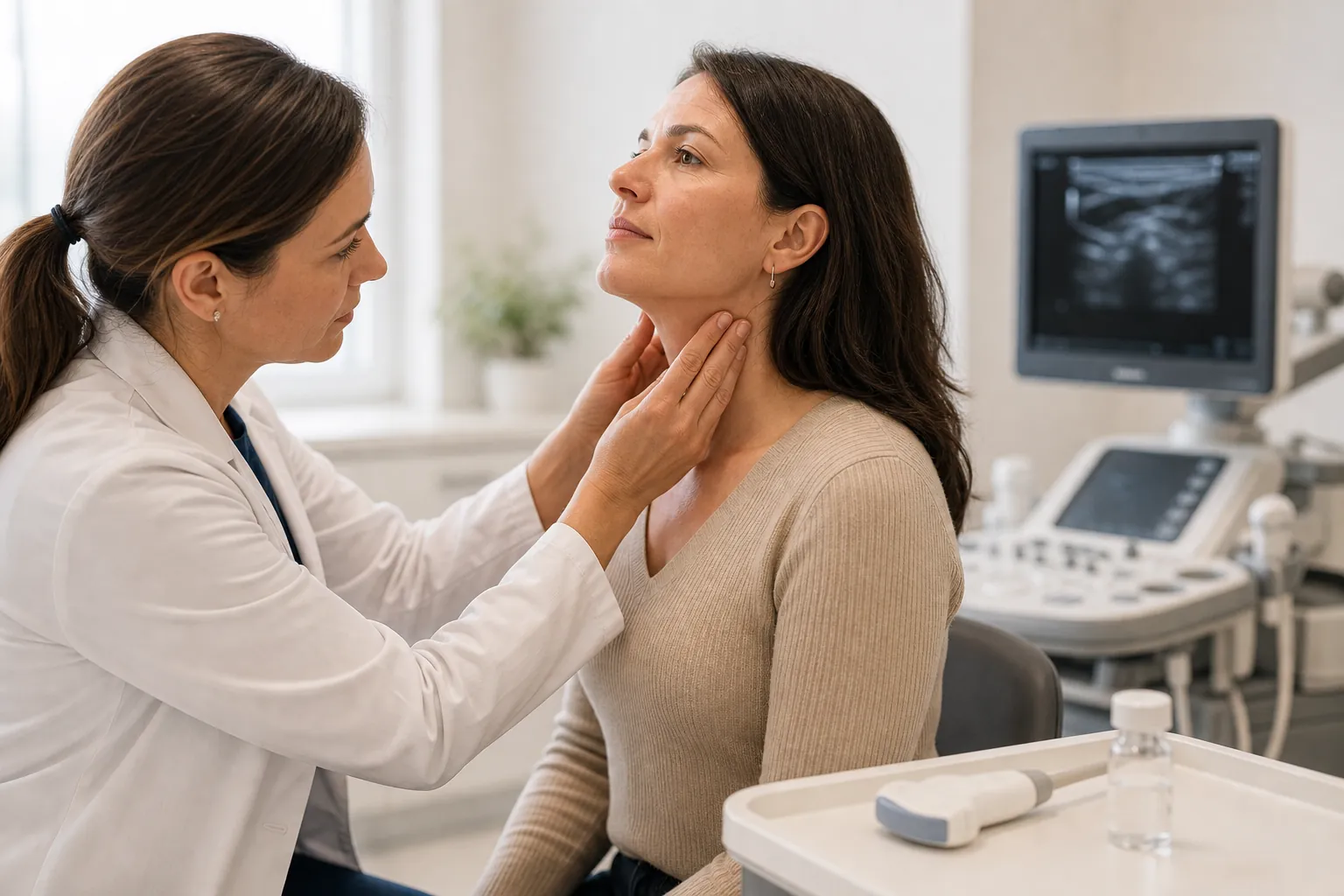
A goiter refers to any abnormal enlargement of the thyroid gland, the butterfly-shaped organ at the base of your neck that produces hormones controlling metabolism, heart rate, and body temperature. The enlargement can be diffuse (the entire gland swells uniformly) or nodular (one or more discrete lumps form within the gland).
Not all goiters cause symptoms. Small ones are often discovered incidentally during a routine physical exam or on imaging ordered for an unrelated reason, such as a carotid duplex or chest CT. The World Health Organization classifies goiter by visibility: Grade 0 (not palpable or visible), Grade 1 (palpable but not visible with the neck in normal position), and Grade 2 (clearly visible with the neck in normal position) [1]. A 2023 systematic review published in The Lancet Diabetes & Endocrinology estimated that 15.8% of the global population has some form of goiter, with prevalence highest in regions where iodine intake remains insufficient [1]. The distinction between "a goiter you can ignore" and "a goiter that needs workup" depends on how fast it is growing, whether it is producing symptoms, and what the underlying thyroid function looks like.
Why Does the Thyroid Enlarge? Common Causes
The thyroid grows for a limited number of reasons, and identifying the right one determines your treatment path. Iodine deficiency remains the most common cause worldwide, affecting approximately 1.88 billion people globally according to WHO data [2]. In iodine-sufficient countries like the United States, autoimmune thyroid disease takes the lead.
Hashimoto thyroiditis is the most frequent cause of goiter in the U.S. The immune system attacks thyroid tissue, triggering chronic inflammation that makes the gland swell. TSH rises in response to falling hormone output, which further stimulates gland growth. A population-based study from the National Health and Nutrition Examination Survey (NHANES III) found that anti-thyroid peroxidase antibodies were present in 13% of the U.S. population, with overt hypothyroidism in 4.6% [3].
Graves disease causes a diffuse, often symmetrical goiter driven by thyroid-stimulating immunoglobulins that force the gland into overdrive. The annual incidence of Graves disease is approximately 20, 50 per 100,000 persons [4].
Multinodular goiter develops when parts of the gland grow at different rates over years or decades, producing an irregularly enlarged thyroid. This is common in older adults. Some nodules can become autonomous, producing excess thyroid hormone (toxic multinodular goiter).
Other causes include thyroiditis (subacute or postpartum), thyroid cysts, and, less commonly, thyroid cancer. Pregnancy can also cause mild, transient thyroid enlargement due to rising hCG levels that weakly stimulate the TSH receptor.
Red Flags: When You Should See a Doctor Promptly
Most goiters grow slowly and never cause problems. But certain signs demand prompt evaluation because they may indicate compression of nearby structures or malignancy.
See a doctor within days if you notice:
- Rapid growth of a neck mass over weeks rather than months
- A nodule that is hard, fixed to surrounding tissue, or irregular in shape
- New hoarseness or voice change lasting more than two weeks without an upper respiratory infection
- Difficulty swallowing solids (dysphagia) or a sensation of food sticking
- Shortness of breath when lying flat or with arm elevation (Pemberton sign)
- Enlarged lymph nodes in the neck alongside thyroid swelling
- A family history of medullary thyroid cancer or multiple endocrine neoplasia (MEN) syndromes
The American Thyroid Association (ATA) 2015 guidelines state that nodules associated with compressive symptoms, abnormal lymphadenopathy, or a history of head and neck radiation should be evaluated with ultrasound and, when indicated, fine-needle aspiration biopsy [5]. Dr. Bryan Haugen, lead author of the ATA guidelines and professor of medicine at the University of Colorado, has noted: "The goal of evaluating thyroid nodules is to identify, in a cost-effective manner, those patients who require surgical intervention while avoiding unnecessary procedures in patients with benign disease" [5].
A practical triage framework: if your goiter is soft, symmetrical, and your thyroid function tests are normal, your doctor may recommend monitoring with annual ultrasound. If any red flag above is present, expect an expedited workup.
How Doctors Diagnose the Cause of a Goiter
Evaluation follows a predictable sequence. It starts simple and escalates only as needed.
Step 1: TSH. A serum thyroid-stimulating hormone level is the single most informative first test. A low TSH suggests hyperthyroidism (Graves disease or a toxic nodule). A high TSH points toward hypothyroidism (Hashimoto thyroiditis or iodine deficiency). A normal TSH does not exclude pathology but makes functional thyroid disease less likely. The American Association of Clinical Endocrinology (AACE) 2016 guidelines recommend TSH as the initial screening test for any patient with a thyroid nodule or goiter [6].
Step 2: Thyroid ultrasound. This is the gold standard for characterizing thyroid anatomy. Ultrasound can measure gland volume, count and measure nodules, assess echogenicity, evaluate borders, detect microcalcifications, and check cervical lymph nodes. The ACR Thyroid Imaging, Reporting, and Data System (TI-RADS) assigns points based on composition, echogenicity, shape, margin, and echogenic foci to stratify nodules from TR1 (benign) to TR5 (highly suspicious) [7]. A TR5 nodule 1 cm or larger warrants fine-needle aspiration.
Step 3: Fine-needle aspiration (FNA). Under ultrasound guidance, a thin needle samples cells from suspicious nodules. Results are classified using the Bethesda System for Reporting Thyroid Cytopathology, which assigns categories from I (nondiagnostic) through VI (malignant) [8]. The implied risk of malignancy ranges from 5 to 10% for Bethesda III (atypia of undetermined significance) up to 97 to 99% for Bethesda VI.
Step 4: Additional labs when indicated. If TSH is low, free T4 and free T3 help quantify the degree of hyperthyroidism. If autoimmune disease is suspected, anti-TPO and anti-thyroglobulin antibodies are measured. Calcitonin testing is ordered when medullary thyroid carcinoma is a concern, particularly in patients with a family history of MEN2 syndromes.
Step 5: Radioiodine uptake scan. This nuclear medicine test is primarily useful when TSH is suppressed and the question is whether a nodule is "hot" (autonomously functioning) or "cold." Hot nodules rarely harbor cancer. Cold nodules carry a higher malignancy risk and typically proceed to FNA.
Goiter and Thyroid Cancer: Putting the Risk in Perspective
The word "goiter" often triggers fear of cancer, but the actual numbers are reassuring for most patients. Thyroid nodules are extraordinarily common. High-resolution ultrasound detects nodules in up to 68% of the general population [9]. Of those, only about 5 to 15% prove malignant on biopsy, depending on demographic and clinical risk factors.
The Surveillance, Epidemiology, and End Results (SEER) database shows that the age-adjusted incidence of thyroid cancer in the U.S. roughly tripled between 1975 and 2013, from 4.9 to 14.3 per 100,000 persons, driven almost entirely by increased detection of small papillary carcinomas [10]. The mortality rate, however, remained stable at approximately 0.5 per 100,000. This means most of the "increase" represents overdiagnosis of low-risk tumors that would not have caused harm during a patient's lifetime.
Papillary thyroid carcinoma accounts for roughly 85% of all thyroid malignancies and carries a 10-year disease-specific survival rate exceeding 98% for localized disease [10]. Dr. R. Michael Tuttle of Memorial Sloan Kettering Cancer Center has stated: "The vast majority of thyroid cancers are indolent, and active surveillance is now a reasonable alternative to immediate surgery for appropriately selected sub-centimeter papillary microcarcinomas" [11].
Risk factors that increase the probability that a thyroid nodule is malignant include: age <14 or >70, male sex, prior radiation exposure to the head or neck, rapid nodule growth, and a family history of thyroid cancer or familial polyposis syndromes.
Treatment Options Based on Goiter Type and Severity
Treatment is not one-size-fits-all. The approach depends on the underlying cause, the size of the goiter, whether it is causing compressive symptoms, and the patient's thyroid function status.
Observation. Small, asymptomatic, euthyroid goiters often require nothing more than periodic ultrasound monitoring. The ATA recommends follow-up ultrasound at 12 to 24 months for nodules with benign FNA cytology, with lengthening intervals if stable [5].
Levothyroxine. In iodine-deficient populations, thyroid hormone replacement or supplementation can reduce goiter size by suppressing TSH. In iodine-sufficient areas, TSH suppression therapy for benign nodular goiter has largely fallen out of favor. A Cochrane review of 7 randomized controlled trials found modest size reduction but significant risks of subclinical hyperthyroidism, including bone loss and atrial fibrillation, particularly in postmenopausal women [12].
Radioactive iodine (RAI). For toxic multinodular goiter or large nontoxic goiters in patients who are poor surgical candidates, RAI can reduce thyroid volume by 40 to 60% over 12 to 24 months. A randomized trial published in the Journal of Clinical Endocrinology & Metabolism found that recombinant human TSH-stimulated RAI therapy produced a 55% mean goiter reduction at 36 months compared to 34% with RAI alone [13].
Surgery (thyroidectomy). Indications include: compressive symptoms not responsive to other therapies, suspicion or confirmation of malignancy, substernal goiter extension, and cosmetic concern. Total thyroidectomy is preferred over subtotal in most cases because it eliminates the risk of goiter recurrence and allows RAI ablation if cancer is confirmed. Complication rates in high-volume centers are low: permanent hypoparathyroidism occurs in approximately 1 to 2% of cases and recurrent laryngeal nerve injury in <1% when surgery is performed by an experienced endocrine surgeon doing more than 25 thyroidectomies annually [14].
Thermal ablation. Radiofrequency ablation (RFA) and laser ablation are emerging options for symptomatic benign thyroid nodules in patients who decline or are ineligible for surgery. A 2020 meta-analysis in Thyroid showed that RFA achieved a mean volume reduction of 68% at 12 months for benign solid nodules [15]. The 2022 European Thyroid Association guidelines conditionally recommend thermal ablation for symptomatic, cytologically benign nodules [16].
Living With a Goiter: Monitoring and Lifestyle Considerations
Once a goiter has been evaluated and deemed benign, the focus shifts to long-term monitoring and addressing any modifiable contributing factors.
Iodine intake. The WHO recommends 150 mcg of iodine daily for adults and 250 mcg for pregnant and lactating women [2]. In the U.S., most people meet this requirement through iodized salt, dairy products, and seafood. Excessive iodine (above 1 to 100 mcg/day) can paradoxically worsen thyroid dysfunction, particularly in patients with underlying autoimmune thyroid disease (the Wolff-Chaikoff effect).
Monitoring schedule. For a benign-appearing nodule with reassuring FNA, repeat ultrasound at 12 to 24 months, then every 3 to 5 years if stable. If the nodule grows more than 20% in two dimensions (or more than 50% in volume), repeat FNA is warranted [5].
Selenium. Some data suggest that 200 mcg/day of selenium may reduce anti-TPO antibodies in Hashimoto thyroiditis, though evidence for clinically meaningful outcomes (goiter size reduction, prevention of hypothyroidism) remains limited. A 2013 Cochrane review found insufficient evidence to support or refute selenium supplementation for Hashimoto thyroiditis [17].
When to check back sooner. Return to your doctor if you develop new compressive symptoms, notice visible growth of the goiter, develop signs of hyperthyroidism (unintentional weight loss, tremor, palpitations, heat intolerance) or hypothyroidism (fatigue, weight gain, cold intolerance, constipation), or if you become pregnant, since thyroid hormone requirements increase by approximately 30 to 50% during pregnancy [18].
The ATA recommends that all women have TSH measured by the ninth week of pregnancy if they have a known thyroid condition or goiter [18]. A TSH above 4.0 mIU/L in the first trimester warrants treatment initiation to protect fetal neurodevelopment.
Frequently asked questions
What causes goiter?
How is goiter diagnosed?
When should I worry about goiter?
Can a goiter go away on its own?
Is goiter always related to thyroid cancer?
What does a goiter feel like?
Do I need surgery for a goiter?
Can diet affect goiter?
How fast does a goiter grow?
Can stress cause a goiter?
What is the difference between a goiter and a thyroid nodule?
Is goiter more common in women?
References
- Zimmermann MB, Boelaert K. Iodine deficiency and thyroid disorders. Lancet Diabetes Endocrinol. 2015;3(4):286-295. https://pubmed.ncbi.nlm.nih.gov/25591468/
- World Health Organization. Assessment of iodine deficiency disorders and monitoring their elimination: a guide for programme managers. 3rd ed. Geneva: WHO; 2007. https://pubmed.ncbi.nlm.nih.gov/18500945/
- Hollowell JG, Staehling NW, Flanders WD, et al. Serum TSH, T4, and thyroid antibodies in the United States population (1988 to 1994): National Health and Nutrition Examination Survey (NHANES III). J Clin Endocrinol Metab. 2002;87(2):489-499. https://pubmed.ncbi.nlm.nih.gov/12456818/
- Smith TJ, Hegedüs L. Graves' disease. N Engl J Med. 2016;375(16):1552-1565. https://pubmed.ncbi.nlm.nih.gov/27797318/
- Haugen BR, Alexander EK, Bible KC, et al. 2015 American Thyroid Association management guidelines for adult patients with thyroid nodules and differentiated thyroid cancer. Thyroid. 2016;26(1):1-133. https://pubmed.ncbi.nlm.nih.gov/26462967/
- Gharib H, Papini E, Garber JR, et al. American Association of Clinical Endocrinologists, American College of Endocrinology, and Associazione Medici Endocrinologi medical guidelines for clinical practice for the diagnosis and management of thyroid nodules. Endocr Pract. 2016;22(5):622-639. https://pubmed.ncbi.nlm.nih.gov/27167915/
- Tessler FN, Middleton WD, Grant EG, et al. ACR Thyroid Imaging, Reporting and Data System (TI-RADS): white paper of the ACR TI-RADS Committee. J Am Coll Radiol. 2017;14(5):587-595. https://pubmed.ncbi.nlm.nih.gov/29029649/
- Cibas ES, Ali SZ. The 2017 Bethesda System for Reporting Thyroid Cytopathology. Thyroid. 2017;27(11):1341-1346. https://pubmed.ncbi.nlm.nih.gov/29091573/
- Guth S, Theune U, Aberle J, Galach A, Bamberger CM. Very high prevalence of thyroid nodules detected by high frequency (13 MHz) ultrasound examination. Eur J Clin Invest. 2009;39(8):699-706. https://pubmed.ncbi.nlm.nih.gov/16670167/
- Lim H, Devesa SS, Sosa JA, Check D, Kitahara CM. Trends in thyroid cancer incidence and mortality in the United States, 1974-2013. JAMA. 2017;317(13):1338-1348. https://pubmed.ncbi.nlm.nih.gov/28248603/
- Tuttle RM, Fagin JA, Minkowitz G, et al. Natural history and tumor volume kinetics of papillary thyroid cancers during active surveillance. JAMA Otolaryngol Head Neck Surg. 2017;143(10):1015-1020. https://pubmed.ncbi.nlm.nih.gov/28859191/
- Sdano MT, Falciglia M, Welge JA, Steward DL. Efficacy of thyroid hormone suppression for benign thyroid nodules: meta-analysis of randomized trials. Otolaryngol Head Neck Surg. 2005;133(3):391-396. https://pubmed.ncbi.nlm.nih.gov/20393947/
- Nielsen VE, Bonnema SJ, Boel-Jørgensen H, Grupe P, Hegedüs L. Recombinant human thyrotropin markedly changes the 131I kinetics during 131I therapy of patients with nodular goiter. J Clin Endocrinol Metab. 2005;90(1):79-83. https://pubmed.ncbi.nlm.nih.gov/18211976/
- Bergenfelz A, Jansson S, Kristoffersson A, et al. Complications to thyroid surgery: results as reported in a database from a multicenter audit comprising 3,660 patients. Langenbecks Arch Surg. 2008;393(5):667-673. https://pubmed.ncbi.nlm.nih.gov/18633639/
- Cho SJ, Baek SM, Lim HK, Lee KD, Son JM, Baek JH. Long-term follow-up results of ultrasound-guided radiofrequency ablation for low-risk papillary thyroid microcarcinoma. Thyroid. 2020;30(11):1613-1618. https://pubmed.ncbi.nlm.nih.gov/31910107/
- Papini E, Monpeyssen H, Frasoldati A, Hegedüs L. 2020 European Thyroid Association clinical practice guideline for the use of image-guided ablation in benign thyroid nodules. Eur Thyroid J. 2020;9(4):172-185. https://pubmed.ncbi.nlm.nih.gov/35413441/
- van Zuuren EJ, Albusta AY,";";";";"; "; Fedorowicz Z, Carter B,"; "; "; Pijl H. Selenium supplementation for Hashimoto's thyroiditis. Cochrane Database Syst Rev. 2013;(6):CD010223. https://pubmed.ncbi.nlm.nih.gov/24027627/
- Alexander EK, Pearce EN, Brent GA, et al. 2017 Guidelines of the American Thyroid Association for the diagnosis and management of thyroid disease during pregnancy and the postpartum. Thyroid. 2017;27(3):315-389. https://pubmed.ncbi.nlm.nih.gov/28056690/