
Thyroid Nodule: What Could Be Causing It
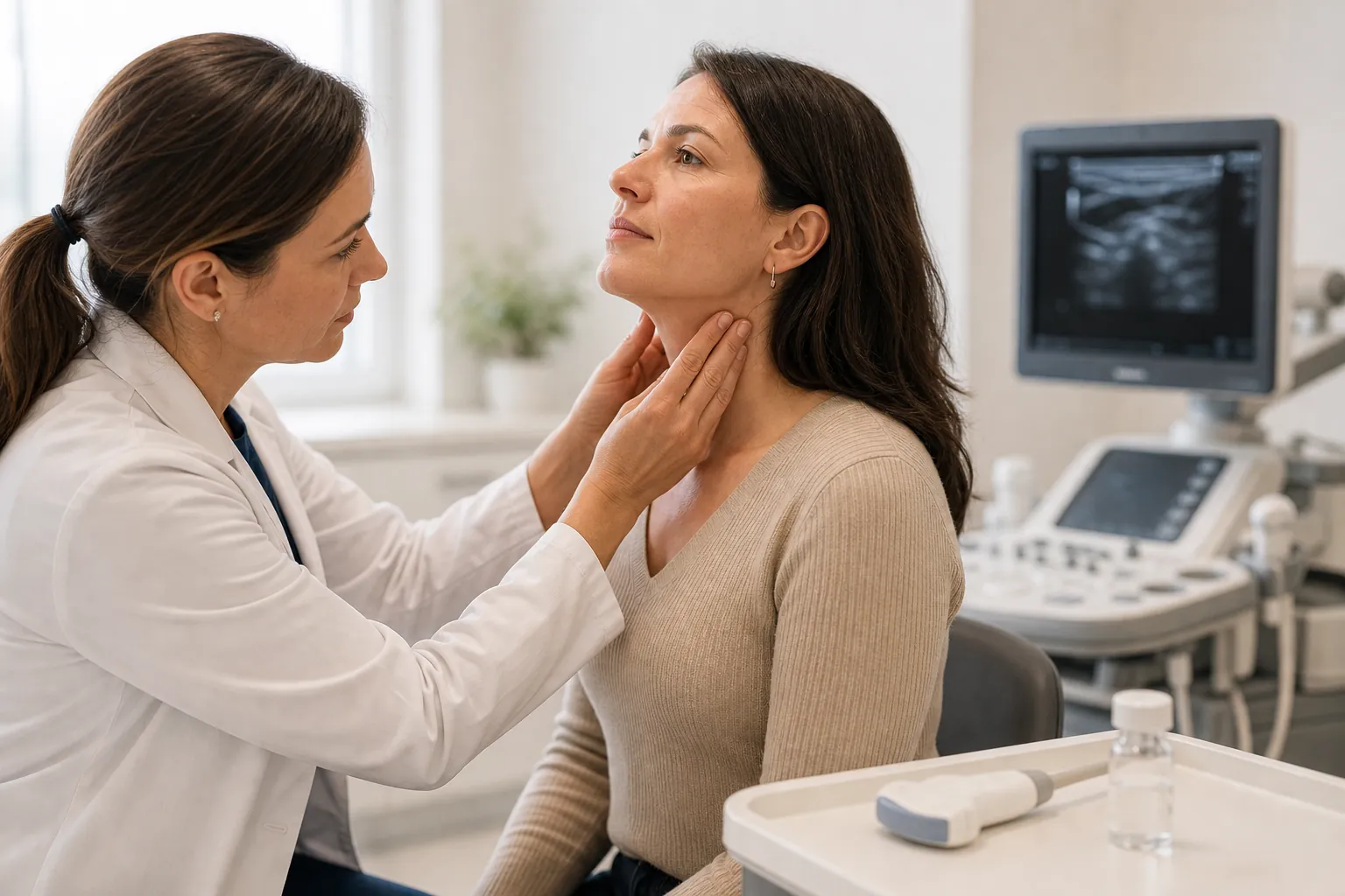
At a glance
- Prevalence / up to 68% of adults have nodules detectable on high-resolution ultrasound
- Benign rate / 90 to 95% of all thyroid nodules are non-cancerous
- Most common cause / colloid (adenomatous) nodules from iodine fluctuation or gland overgrowth
- Cancer risk / approximately 5 to 10% of clinically detected nodules are malignant
- Key diagnostic tool / thyroid ultrasound with TI-RADS scoring
- Biopsy threshold / ATA recommends FNA for nodules 1 cm or larger with suspicious sonographic features
- TSH role / a suppressed TSH suggests autonomous (hot) nodule, which is rarely malignant
- Radiation history / prior head or neck radiation raises cancer risk 2 to 4 fold
- Sex distribution / women are 4 times more likely than men to develop thyroid nodules
- Monitoring interval / benign nodules typically re-imaged at 12 to 24 months
Why Thyroid Nodules Are So Common
Thyroid nodules are among the most frequently encountered endocrine findings in clinical practice. High-resolution ultrasound detects nodules in up to 68% of randomly selected adults, according to a 2010 population study published in Thyroid [1]. Most people never know they have one.
The thyroid gland sits at the base of the neck and produces hormones (T3 and T4) that regulate metabolism, heart rate, and body temperature. Nodules form when clusters of thyroid cells grow abnormally or when fluid-filled cysts develop within the gland. The overwhelming majority are asymptomatic and discovered incidentally during imaging for unrelated conditions, such as carotid duplex ultrasound or CT scans of the chest.
Several factors increase nodule prevalence. Female sex carries a 4:1 risk ratio compared to males [2]. Age matters: the incidence rises roughly 2% per year after age 20. Iodine deficiency, a declining concern in the United States but still prevalent globally, stimulates thyroid-stimulating hormone (TSH) secretion and promotes nodular growth [3]. A family history of thyroid disease, prior radiation exposure to the head or neck (especially in childhood), and chronic autoimmune thyroiditis (Hashimoto disease) each independently raise the probability of finding one or more nodules on examination.
Benign Causes: The 90 to 95 Percent
The vast majority of thyroid nodules fall into a handful of benign categories. Understanding these helps clinicians (and patients) avoid unnecessary anxiety when imaging reveals a lump.
Colloid nodules are the single most common type. They arise when normal thyroid tissue overgrows and fills with stored colloid, the protein-rich substance that serves as a reservoir for thyroid hormone. Colloid nodules are not neoplastic. They grow slowly, rarely cause symptoms, and almost never become malignant. Multinodular goiter, a condition in which the thyroid contains multiple colloid nodules, affects an estimated 5% of the U.S. population [4].
Thyroid cysts are fluid-filled sacs. Purely cystic nodules carry a malignancy risk below 1% [5]. Mixed cystic-solid nodules, where both fluid and tissue components are present, require closer evaluation because the solid portion can occasionally harbor papillary carcinoma.
Follicular adenomas are true benign neoplasms. They grow within a capsule and do not invade surrounding tissue. On fine-needle aspiration (FNA), they can be difficult to distinguish from follicular carcinoma because the distinction depends on capsular or vascular invasion, features visible only on surgical pathology. This diagnostic gray zone accounts for many of the "indeterminate" cytology results that lead to diagnostic lobectomy.
Hashimoto thyroiditis produces chronic lymphocytic infiltration that can create pseudonodules, areas of focal inflammation that mimic discrete nodules on ultrasound. A 2017 meta-analysis in The Journal of Clinical Endocrinology & Metabolism found that Hashimoto thyroiditis patients have a slightly elevated risk of papillary thyroid cancer (odds ratio 1.6), though the absolute risk remains low [6].
Subacute thyroiditis (de Quervain thyroiditis), triggered by viral infection, can produce tender, painful nodules that resolve over weeks to months without intervention. These are self-limiting, though they may temporarily cause thyrotoxicosis followed by hypothyroidism before the gland recovers.
When a Nodule Might Be Cancer
Thyroid cancer accounts for 5 to 10% of all thyroid nodules. The good news: most thyroid cancers are highly treatable. The 5-year survival rate for papillary thyroid carcinoma, the most common subtype (representing about 80% of thyroid malignancies), exceeds 98% for localized disease [7].
Four histological types dominate. Papillary carcinoma is the most frequent and carries the best prognosis. Follicular carcinoma accounts for 10 to 15% of cases and tends to spread hematogenously rather than via lymph nodes. Medullary thyroid carcinoma (MTC) originates from parafollicular C cells, produces calcitonin, and can be sporadic or hereditary (MEN2 syndrome). Anaplastic carcinoma is rare (1 to 2% of thyroid cancers) but aggressive, with a median survival of 5 months [8].
Risk factors that raise suspicion for malignancy include a history of childhood head or neck irradiation, a family history of thyroid cancer or MEN2 syndrome, rapid nodule growth, fixation to surrounding structures, hoarseness from vocal cord paralysis, and ipsilateral cervical lymphadenopathy. Male sex, while less commonly associated with nodules overall, carries a higher per-nodule cancer risk compared to female sex [9].
The 2015 ATA guidelines provide a risk-stratified framework for evaluating nodules based on ultrasound pattern [10]. Nodules with microcalcifications, irregular margins, taller-than-wide shape, or marked hypoechogenicity fall into the "high suspicion" category (estimated malignancy risk above 70%) and warrant FNA at sizes as small as 1 cm. Spongiform or purely cystic nodules, by contrast, are classified as "very low suspicion" (malignancy risk <3%) and may be observed without biopsy even at larger sizes.
How Thyroid Nodules Are Diagnosed
Diagnosis follows a structured pathway: clinical assessment, blood work, imaging, and (when indicated) tissue sampling. Each step narrows the differential.
Physical examination detects only about 4 to 7% of nodules, typically those larger than 1.5 cm or located anteriorly in the gland [11]. Palpation cannot reliably distinguish benign from malignant. A hard, fixed nodule with associated lymphadenopathy raises concern, but soft, mobile nodules can also harbor cancer.
TSH measurement is the first laboratory test. A low TSH suggests the nodule may be autonomously producing thyroid hormone (a "hot" nodule), which is rarely malignant. If TSH is suppressed, a radioiodine (I-123) uptake scan can confirm autonomous function. Hot nodules on scintigraphy have a cancer risk below 1%, and FNA is generally not indicated [10]. A normal or elevated TSH does not rule out malignancy but shifts the diagnostic pathway toward ultrasound-guided evaluation.
Thyroid ultrasound is the single most informative imaging modality. The American College of Radiology's Thyroid Imaging Reporting and Data System (TI-RADS) assigns points based on composition, echogenicity, shape, margin characteristics, and echogenic foci [12]. The composite score determines whether FNA or surveillance is recommended.
According to ACR TI-RADS:
- TR1 (benign): no FNA needed
- TR2 (not suspicious): no FNA needed
- TR3 (mildly suspicious): FNA if 2.5 cm or larger
- TR4 (moderately suspicious): FNA if 1.5 cm or larger
- TR5 (highly suspicious): FNA if 1 cm or larger
Fine-needle aspiration biopsy is the definitive presurgical test. A 2019 review in The Lancet Diabetes & Endocrinology reported that FNA has a sensitivity of 89 to 98% and specificity of 92 to 100% for detecting thyroid malignancy when an adequate sample is obtained [13]. Cytology results are reported using the Bethesda System for Reporting Thyroid Cytopathology, which assigns nodules to one of six categories, each with an implied cancer risk:
- Bethesda I (nondiagnostic): 5 to 10% malignancy risk; repeat FNA recommended
- Bethesda II (benign): 0 to 3% risk; clinical follow-up
- Bethesda III (atypia of undetermined significance): 6 to 18% risk
- Bethesda IV (follicular neoplasm): 10 to 40% risk
- Bethesda V (suspicious for malignancy): 45 to 60% risk
- Bethesda VI (malignant): 94 to 96% risk [14]
Molecular testing has changed diagnostic decision-making for indeterminate nodules (Bethesda III and IV). The Afirma Gene Sequencing Classifier and ThyroSeq v3 genomic panel help determine whether a nodule with ambiguous cytology can be safely surveilled or requires surgery. ThyroSeq v3 demonstrated a sensitivity of 94% and specificity of 82% in a 2018 validation study published in JAMA Oncology [15]. Dr. Yuri Nikiforov, who led that study, stated: "Molecular testing allows us to reduce unnecessary surgeries by about 50% in patients with indeterminate cytology results."
Treatment Options by Diagnosis
Treatment depends entirely on whether the nodule is benign, indeterminate, or malignant. No single approach fits every case.
Benign nodules (Bethesda II) require no treatment in most cases. The ATA recommends repeat ultrasound at 12 to 24 months. If the nodule grows by more than 50% in volume (or 20% in at least two dimensions with a minimum increase of 2 mm), repeat FNA is warranted [10]. Patients with large benign nodules causing compressive symptoms (dysphagia, dyspnea, cosmetic concern) may benefit from thyroid lobectomy, radiofrequency ablation (RFA), or ethanol injection for cystic nodules.
Radiofrequency ablation has gained traction as a non-surgical option for symptomatic benign nodules. A 2020 Korean multicenter study (N=450) published in Radiology showed that RFA reduced nodule volume by a mean of 68.4% at 1 year, with a major complication rate of 1.4% [16]. The ATA has not yet incorporated RFA into formal guidelines, but the Korean Society of Thyroid Radiology and the European Thyroid Association both endorse it for selected cases.
Indeterminate nodules (Bethesda III/IV) present a clinical decision point. Options include repeat FNA, molecular testing, or diagnostic lobectomy. Molecular testing has reduced the rate of diagnostic surgery for Bethesda III nodules from roughly 65% to about 30% in centers that routinely use genomic classifiers [15]. When molecular results are suspicious, lobectomy with possible completion thyroidectomy is the standard approach.
Malignant nodules are treated primarily with surgery. For papillary carcinomas larger than 4 cm, with extrathyroidal extension, or with distant metastases, total thyroidectomy followed by radioactive iodine (RAI) ablation remains the standard of care. For papillary microcarcinomas (1 cm or smaller) without aggressive features, the 2015 ATA guidelines introduced active surveillance as an alternative to immediate surgery, citing data from Kuma Hospital in Japan where 1,235 patients with low-risk papillary microcarcinoma were followed for a mean of 5 years. Only 8% showed tumor growth exceeding 3 mm, and none developed distant metastases [17].
Dr. R. Michael Tuttle of Memorial Sloan Kettering Cancer Center has commented on this approach: "Active surveillance for low-risk papillary microcarcinoma is not doing nothing. It is a structured management strategy with defined follow-up intervals and clear intervention triggers."
Autonomous (hot) nodules that cause hyperthyroidism can be managed with radioactive iodine ablation, surgical excision, or antithyroid medications (methimazole) as a bridge to definitive treatment. RAI is the most commonly used definitive therapy in the United States for toxic adenomas [18].
Thyroid Nodules in Special Populations
Certain groups require modified evaluation and management strategies.
Pregnant patients present a unique challenge. Thyroid nodules discovered during pregnancy should be evaluated with ultrasound and TSH measurement, but radioiodine scanning is contraindicated. FNA can be performed safely during any trimester. If cytology reveals malignancy, surgery (preferably in the second trimester) is recommended for aggressive histologies. Low-risk papillary carcinoma diagnosed during pregnancy can often be monitored until after delivery without affecting oncologic outcomes, as the ATA 2015 guidelines note [10].
Children and adolescents have a higher per-nodule malignancy rate (22 to 26%) compared to adults [19]. The ATA published separate pediatric guidelines in 2015, recommending FNA for nodules 1 cm or larger with suspicious sonographic features and a lower threshold for surgical intervention given the higher cancer prevalence in this age group.
Patients with prior radiation exposure, including survivors of childhood cancers treated with external beam radiation to the head, neck, or upper mediastinum, carry a 2 to 4 fold increased risk of thyroid malignancy. Screening ultrasound beginning 5 years after radiation exposure is recommended by the Children's Oncology Group [20].
Elderly patients require careful risk-benefit analysis. While thyroid cancer incidence peaks in middle age, anaplastic carcinoma occurs predominantly in patients over 60. Conversely, aggressive workup and surgery in frail elderly patients may carry more risk than the nodule itself. Shared decision-making is essential.
Monitoring and Long-Term Follow-Up
Benign thyroid nodules do not disappear. They persist, may grow slowly, and occasionally require re-evaluation. The ATA recommends the following surveillance schedule for cytologically benign nodules:
- High-suspicion ultrasound pattern: repeat ultrasound at 12 months
- Low-to-intermediate suspicion pattern: repeat ultrasound at 12 to 24 months
- Very low suspicion or spongiform: repeat ultrasound at 24 months or longer
If two consecutive ultrasounds show stability (no significant growth), the interval can be extended. Nodules that grow should undergo repeat FNA. The false-negative rate of a single benign FNA is approximately 3%, but drops below 1% after two concordant benign results [10].
Patients should be counseled that thyroid nodules are overwhelmingly benign and that incidental discovery does not indicate disease. A 2020 cross-sectional study in Thyroid found that 37% of patients with benign nodules reported moderate to severe anxiety about their diagnosis, a finding that argues for clear communication during initial workup [21]. Clinicians who proactively explain the low malignancy risk and structured surveillance plan reduce patient distress and unnecessary repeat testing.
Suppressive levothyroxine therapy (lowering TSH to reduce nodule growth) was once common practice but is no longer recommended by the ATA for benign nodules in iodine-sufficient populations. A 2014 Cochrane review found no significant long-term benefit of TSH suppression for nodule volume reduction and documented increased risks of atrial fibrillation and bone loss [22].
Annual TSH monitoring remains appropriate for patients with nodules and coexisting Hashimoto thyroiditis, given the progressive nature of autoimmune thyroid destruction and the eventual need for levothyroxine replacement in many of these patients.
Frequently asked questions
What causes thyroid nodules?
How is a thyroid nodule diagnosed?
When should I worry about a thyroid nodule?
Can thyroid nodules go away on their own?
Do all thyroid nodules need a biopsy?
What is the difference between a hot and cold thyroid nodule?
Is thyroid nodule surgery always necessary?
Can stress cause thyroid nodules?
How fast do thyroid nodules grow?
Are thyroid nodules hereditary?
What does an indeterminate thyroid biopsy result mean?
Can you live with a thyroid nodule without treatment?
References
- Guth S, Theune U, Aberle J, Galach A, Bamberger CM. Very high prevalence of thyroid nodules detected by high frequency (13 MHz) ultrasound examination. Eur J Clin Invest. 2009;39(8):699-706. https://pubmed.ncbi.nlm.nih.gov/19601965/
- Dean DS, Gharib H. Epidemiology of thyroid nodules. Best Pract Res Clin Endocrinol Metab. 2008;22(6):901-911. https://pubmed.ncbi.nlm.nih.gov/19041821/
- Zimmermann MB, Boelaert K. Iodine deficiency and thyroid disorders. Lancet Diabetes Endocrinol. 2015;3(4):286-295. https://pubmed.ncbi.nlm.nih.gov/25591468/
- Burman KD, Wartofsky L. Thyroid nodules. N Engl J Med. 2015;373(24):2347-2356. https://pubmed.ncbi.nlm.nih.gov/26650154/
- Brito JP, Yarur AJ, Engber TM, et al. Prevalence of thyroid cancer in multinodular goiter versus single nodules: a systematic review. Thyroid. 2013;23(4):449-455. https://pubmed.ncbi.nlm.nih.gov/23067375/
- Resende de Paiva C, Grønhøj C, Feldt-Rasmussen U, von Buchwald C. Association between Hashimoto thyroiditis and thyroid cancer: a systematic review and meta-analysis. J Clin Endocrinol Metab. 2017;102(9):3116-3121. https://pubmed.ncbi.nlm.nih.gov/28609832/
- Surveillance, Epidemiology, and End Results Program. Cancer stat facts: thyroid cancer. National Cancer Institute. https://www.ncbi.nlm.nih.gov/books/NBK459289/
- Molinaro E, Romei C, Biagini A, et al. Anaplastic thyroid carcinoma: from clinicopathology to genetics and advanced therapies. Nat Rev Endocrinol. 2017;13(11):644-660. https://pubmed.ncbi.nlm.nih.gov/28707679/
- Brito JP, Morris JC, Montori VM. Thyroid cancer: zealous imaging has increased detection and treatment of low risk tumours. BMJ. 2013;347:f4706. https://pubmed.ncbi.nlm.nih.gov/23982465/
- Haugen BR, Alexander EK, Bible KC, et al. 2015 American Thyroid Association management guidelines for adult patients with thyroid nodules and differentiated thyroid cancer. Thyroid. 2016;26(1):1-133. https://pubmed.ncbi.nlm.nih.gov/26462967/
- Brander A, Viikinkoski P, Nickels J, Kivisaari L. Thyroid gland: US screening in a random adult population. Radiology. 1991;181(3):683-687. https://pubmed.ncbi.nlm.nih.gov/1947082/
- Tessler FN, Middleton WD, Grant EG, et al. ACR Thyroid Imaging, Reporting and Data System (TI-RADS): white paper of the ACR TI-RADS Committee. J Am Coll Radiol. 2017;14(5):587-595. https://pubmed.ncbi.nlm.nih.gov/28372962/
- Cibas ES, Ali SZ. The 2017 Bethesda System for Reporting Thyroid Cytopathology. Thyroid. 2017;27(11):1341-1346. https://pubmed.ncbi.nlm.nih.gov/29091573/
- Ali SZ, Cibas ES. The Bethesda System for Reporting Thyroid Cytopathology. 2nd ed. Springer; 2018. https://pubmed.ncbi.nlm.nih.gov/29091573/
- Steward DL, Carty SE, Sippel RS, et al. Performance of a multigene genomic classifier in thyroid nodules with indeterminate cytology: a prospective blinded multicenter study. JAMA Oncol. 2019;5(2):204-212. https://pubmed.ncbi.nlm.nih.gov/30419129/
- Baek JH, Lee JH, Sung JY, et al. Complications encountered in the treatment of benign thyroid nodules with US-guided radiofrequency ablation: a multicenter study. Radiology. 2012;262(1):335-342. Complications encountered in the treatment of benign thyroid nodules with US-guided radiofrequency ablation: a multicenter study
- Ito Y, Miyauchi A, Kihara M, Higashiyama T, Kobayashi K, Miya A. Patient age is significantly related to the progression of papillary microcarcinoma of the thyroid under observation. Thyroid. 2014;24(1):27-34. https://pubmed.ncbi.nlm.nih.gov/24001104/
- Ross DS, Burch HB, Cooper DS, et al. 2016 American Thyroid Association guidelines for diagnosis and management of hyperthyroidism and other causes of thyrotoxicosis. Thyroid. 2016;26(10):1343-1421. https://pubmed.ncbi.nlm.nih.gov/27521067/
- Francis GL, Waguespack SG, Bauer AJ, et al. Management guidelines for children with thyroid nodules and differentiated thyroid cancer. Thyroid. 2015;25(7):716-759. https://pubmed.ncbi.nlm.nih.gov/25900731/
- Children's Oncology Group. Long-term follow-up guidelines for survivors of childhood, adolescent, and young adult cancers. Version 5.0. 2018. https://www.ncbi.nlm.nih.gov/books/NBK543609/
- Papaleontiou M, Reyes-Gastelum D, Gay BL, et al. Worry in thyroid cancer survivors with a favorable prognosis. Thyroid. 2019;29(8):1080-1088. Worry in Thyroid Cancer Survivors with a Favorable Prognosis
- Sdano MT, Falciglia M, Welge JA, Steward DL. Efficacy of thyroid hormone suppression for benign thyroid nodules: meta-analysis of randomized trials. Otolaryngol Head Neck Surg. 2005;133(3):391-396. Efficacy of thyroid hormone suppression for benign thyroid nodules: meta-analysis of randomized trials