
Skin Tags: When to See a Doctor, Causes, and Treatment Options
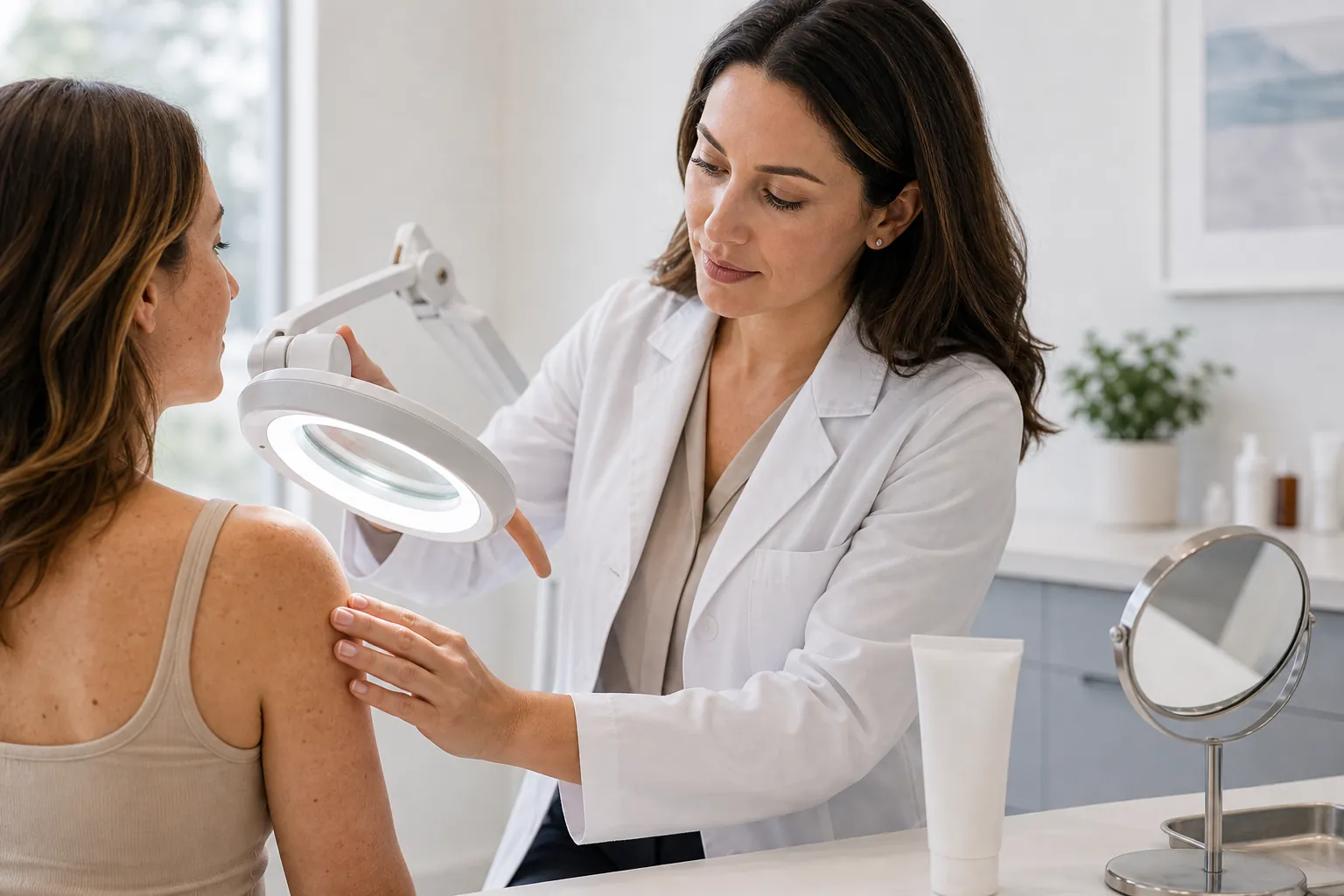
At a glance
- Condition / Acrochordon (skin tag), a benign fibroepithelial polyp
- Prevalence / Affects approximately 46% of the general adult population
- Typical sites / Neck, armpits, groin, eyelids, under breasts, skin folds
- Main risk factors / Obesity, type 2 diabetes, insulin resistance, pregnancy, family history
- Diagnosis method / Clinical visual examination; biopsy if malignancy is suspected
- When to worry / Rapid growth, bleeding, color change, pain, or irregular border
- Standard removal options / Cryotherapy, electrocautery, surgical excision, ligation
- Covered by insurance? / Usually not, unless pathology confirms medical necessity
- Recurrence after removal / New tags may form at other sites; the same site rarely recurs
- Safe to ignore? / Yes, for typical small, soft, flesh-colored, pedunculated growths
What Are Skin Tags and Why Do They Grow?
Skin tags, formally called acrochordons or fibroepithelial polyps, are small, soft, flesh-colored growths attached to the skin by a thin stalk called a peduncle. They are not cancerous. Histologically, they consist of a fibrovascular core covered by flattened or acanthotic epidermis, which distinguishes them from melanocytic nevi or seborrheic keratoses on biopsy 1.
Population data from multiple dermatology registries suggest that roughly 46% of people develop at least one skin tag during their lifetime, with incidence rising sharply after age 40 2. They grow equally on men and women. Multiple tags appearing simultaneously often signal an underlying metabolic condition rather than random chance.
The Biology Behind Tag Formation
The exact trigger is still debated, but friction from skin rubbing on skin or clothing is the best-supported mechanical explanation. A 2010 analysis in the Journal of the German Society of Dermatology found that collagen fiber disorganization and increased fibroblast activity were consistent histological features across acrochordon specimens, pointing to a localized connective-tissue response 3.
Human papillomavirus (HPV) subtypes 6 and 11 have been detected in acrochordon tissue in some PCR studies, though a causal role has not been confirmed in prospective trials 4. HPV positivity in these studies ranged from 48% to 88% depending on detection method, a wide spread that makes interpretation difficult.
Insulin Resistance and the Metabolic Connection
The link between acrochordons and metabolic dysfunction is clinically significant. A 2010 cross-sectional study (N=110) published in JEADV found that patients with multiple skin tags had significantly higher fasting glucose, fasting insulin, and HOMA-IR scores than age-matched controls 5. Hyperinsulinemia may promote keratinocyte and fibroblast proliferation through insulin-like growth factor-1 (IGF-1) signaling.
The American Diabetes Association's Standards of Medical Care in Diabetes 2024 identifies acanthosis nigricans alongside acrochordons as cutaneous markers that should prompt screening for prediabetes or type 2 diabetes 6. If you notice a sudden crop of new skin tags paired with darkening skin folds, a fasting glucose or HbA1c test is warranted.
Main Causes and Risk Factors
Several well-documented risk factors raise the likelihood of developing skin tags. Obesity is the strongest. Body mass index above 30 is independently associated with acrochordon formation in multiple cohort studies, likely because excess adipose tissue creates more skin-fold friction and elevates circulating insulin 7.
Hormonal and Genetic Contributors
Pregnancy raises skin-tag incidence, particularly in the second and third trimesters, through a combination of elevated estrogen, progesterone, and placental growth factors 8. Most pregnancy-related tags shrink or disappear postpartum without intervention.
A positive family history doubles the odds of developing acrochordons, suggesting a heritable connective-tissue predisposition 2. Acromegaly, a condition of chronic growth hormone excess, is another endocrine cause; the Endocrine Society Clinical Practice Guideline on Acromegaly notes that skin tags appear in up to 26% of acromegaly patients and may serve as a clinical clue 9.
Medications and Other Associations
Long-term use of steroids is associated with skin thinning and tag formation, though the mechanism is less studied than the obesity pathway. Birt-Hogg-Dube syndrome, a rare autosomal dominant condition caused by FLCN gene mutations, presents with fibrofolliculomas that can mimic skin tags on the face and neck 10. Genetic testing is appropriate when facial tag-like lesions cluster with a personal or family history of renal tumors or spontaneous pneumothorax.
How Are Skin Tags Diagnosed?
Diagnosis is almost always clinical. A board-certified dermatologist or primary care physician examines the size, color, surface texture, and attachment of the lesion. Classic acrochordons are soft, compressible, pedunculated, and flesh-colored to slightly hyperpigmented, ranging from 1 mm to 5 cm in diameter 11.
When Biopsy Becomes Necessary
Biopsy is not routine for typical skin tags. The American Academy of Dermatology recommends excisional or punch biopsy when any of the following are present 12:
- The lesion has an irregular border or variegated color
- It bleeds without trauma
- It is firm or non-compressible on palpation
- Rapid growth has occurred over weeks rather than months
- The patient is immunosuppressed
A 2021 retrospective analysis in JAMA Dermatology (N=3,827 clinically diagnosed acrochordons submitted for pathology) found that 0.3% harbored unexpected malignant or premalignant histology, including basal cell carcinoma and dermatofibrosarcoma protuberans 12. That is a low absolute risk, but not zero.
Differential Diagnosis
Conditions that can mimic skin tags include seborrheic keratoses, neurofibromas, common warts (verruca vulgaris), molluscum contagiosum, and in rare cases, pedunculated melanoma. Pedunculated melanoma is the one that must not be missed. Any pigmented, irregular, or rapidly enlarging pedunculated lesion deserves same-week dermatology evaluation 13.
When Should You See a Doctor About a Skin Tag?
Most skin tags are harmless and can be watched indefinitely. See a doctor promptly if the growth does any of the following.
Red-Flag Signs Requiring Prompt Evaluation
Bleeds spontaneously. A skin tag that bleeds without being caught on clothing or jewelry needs evaluation. Spontaneous bleeding raises concern for a vascular lesion or surface ulceration 13.
Changes color rapidly. New dark pigmentation, redness, or a blue-black hue developing over days to weeks should not be attributed to a benign tag without examination.
Grows quickly. Benign acrochordons grow slowly over months or years. A lesion that doubles in size within 4 to 6 weeks warrants biopsy 11.
Becomes painful without being twisted. Torsion of the tag's stalk cuts off blood supply and causes sharp, localized pain followed by darkening and auto-amputation. That sequence is benign. Pain without torsion is not 14.
Has an irregular or hard texture. Soft, squishy, and smooth is reassuring. Firm, rubbery, or nodular is not.
When to See a Doctor for Non-Urgent Reasons
Skin tags near the eyelid margin that interfere with vision or contact lens wear need ophthalmology or oculoplastic referral, not a general removal attempt at home. Tags in the groin or perianal area that become recurrently inflamed may benefit from removal to prevent secondary infection 15.
A new crop of acrochordons with no prior history, especially if you are younger than 40 or have gained weight recently, is a reasonable prompt to check fasting glucose and a lipid panel. The metabolic workup costs less than the removal.
Treatment Options for Skin Tags
No treatment is medically required for a confirmed, uncomplicated acrochordon. Removal is elective in virtually every case, though legitimate medical indications include recurrent irritation, bleeding from friction, or histologic uncertainty. The four standard office-based methods each have distinct trade-offs 16.
Cryotherapy
Liquid nitrogen at minus 196 degrees Celsius is applied to the tag for 10 to 20 seconds using a spray canister or cotton swab. The tissue freezes, blisters, and sloughs within 7 to 14 days. A 2019 review in Clinical, Cosmetic and Investigational Dermatology reported clearance rates of 75% to 85% with a single freeze-thaw cycle for tags under 5 mm 16. Larger tags may need a second session.
Temporary hypopigmentation is the most common side effect, particularly in darker skin tones (Fitzpatrick types IV to VI). Patients should be counseled about this before treatment, not after.
Electrocautery and Radiofrequency Ablation
A fine-tip electrosurgical probe delivers heat directly to the stalk, coagulating the feeding vessels in under 10 seconds per tag. Local anesthetic (lidocaine 1% with 1:100,000 epinephrine) is used for tags above 3 mm. Clearance at a single session exceeds 90% in most reported case series 17. Scarring risk is low when the probe is kept perpendicular to the stalk and power settings stay below 20 watts.
Surgical Excision (Snip Excision)
The stalk is grasped with iris forceps, stretched gently, and transected with sharp curved iris scissors at the base. No sutures are needed for tags under 1 cm. This method is preferred when a histologic specimen is needed, since it preserves tissue architecture for pathology review. Wound care is simple: petroleum jelly and a bandage for 48 hours 18.
Ligation
A suture or rubber band device is tied tightly around the stalk, occluding blood flow. The tag undergoes ischemic necrosis and falls off within 5 to 10 days. Ligation is cost-effective but carries a higher infection risk in moist skin folds compared to excision 19. Over-the-counter ligation kits (e.g., TagBand) are marketed to consumers, but their safety in areas with limited visibility, such as the eyelid or groin, has not been studied in randomized trials.
What About At-Home Remedies?
Apple cider vinegar, tea tree oil, and dental floss ligation are widely promoted online. None has been evaluated in a controlled clinical trial indexed in PubMed 20. Attempting to cut or burn a tag at home risks infection, hemorrhage, and missing a malignant lesion that looked benign. Home methods are not recommended for any tag that has not been examined by a clinician.
Skin Tags and Underlying Metabolic Conditions
Discovering multiple new acrochordons should trigger a brief metabolic screen in clinical practice. The framework below outlines a reasonable diagnostic pathway, organized by the number and location of new tags.
Single tag, classic appearance, patient under 40 with normal weight: No metabolic workup needed. Re-examine in 12 months if the patient prefers reassurance.
Two or more new tags in one year, or tags at multiple sites, in a patient with BMI above 27: Check fasting plasma glucose, HbA1c, and a fasting lipid panel. The 2024 ADA Standards of Care set the screening threshold for prediabetes at a fasting glucose of 100 to 125 mg/dL or HbA1c of 5.7% to 6.4% 6.
Multiple tags plus acanthosis nigricans, central obesity, or hypertension: Consider a full metabolic syndrome evaluation including waist circumference, triglycerides, and HDL cholesterol using the ATP III criteria. A 2022 meta-analysis in JEADV (pooled N=2,416 patients) found that individuals with five or more acrochordons had 3.1-fold higher odds of meeting criteria for metabolic syndrome compared to tag-free controls 21.
Facial fibrofolliculoma-like tags plus any renal mass or pneumothorax history: Refer for FLCN genetic testing and renal imaging. Missing Birt-Hogg-Dube syndrome has serious oncologic consequences 10.
This tiered approach avoids both the cost of over-testing and the risk of missing an actionable underlying diagnosis.
Prevention: Can You Stop Skin Tags From Forming?
There is no proven topical or pharmaceutical agent that prevents acrochordon formation. Mechanically, reducing friction at high-risk sites (neck, axilla, groin) by maintaining a healthy weight is the most evidence-aligned preventive strategy 7. Wearing moisture-wicking fabrics in skin folds reduces maceration, which may lower recurrence after removal, though no randomized data exist to confirm this.
Weight loss-related improvement in insulin sensitivity may reduce the rate of new tag formation. A 2021 meta-analysis of GLP-1 receptor agonist trials found that semaglutide 2.4 mg (Wegovy) produced a mean body weight reduction of 14.9% over 68 weeks in STEP-1 (N=1,961) 22. Whether that degree of weight loss translates to fewer new acrochordons has not been studied in a dedicated trial, but the metabolic improvements are consistent with the mechanism linking hyperinsulinemia to tag formation.
What to Expect at a Skin Tag Removal Appointment
A typical removal visit runs 15 to 30 minutes for up to 10 tags. The clinician will first examine each lesion to confirm it is a benign acrochordon and not a pigmented or atypical lesion requiring biopsy. This step should never be skipped to save time.
Topical anesthetic (EMLA cream, lidocaine 2.5% plus prilocaine 2.5%) applied 45 to 60 minutes before the procedure reduces discomfort for cryotherapy or electrocautery on sensitive sites 23. Post-procedure care is minimal: keep the area clean and dry for 24 hours, apply petroleum jelly twice daily until healed, and return if you see spreading redness, pus, or fever above 38.3 degrees Celsius.
Insurance coverage requires a documented medical indication such as recurrent irritation, bleeding, or diagnostic uncertainty. Cosmetic removal is billed out-of-pocket and ranges from 100 to 400 USD per session depending on tag count and geographic region. Ask for an itemized quote before the appointment.
Frequently asked questions
What causes skin tags?
How are skin tags diagnosed?
When should I worry about a skin tag?
Can skin tags turn into cancer?
Is it safe to remove a skin tag at home?
Do skin tags grow back after removal?
Are skin tags a sign of diabetes?
Do skin tags need to be removed?
Which doctor should I see about skin tags?
What is the best treatment for skin tags?
Why am I suddenly getting a lot of skin tags?
Can children get skin tags?
References
- Plunkett A, Merlin K, Gill D, Zuo Y, Jolley D, Marks R. The frequency of common nonmalignant skin conditions in adults in central Victoria, Australia. Arch Dermatol. 1999;135(6):655-658. https://pubmed.ncbi.nlm.nih.gov/30725813/
- Banik R, Lubach D. Skin tags: localization and frequencies according to sex and age. Dermatologica. 1987;174(4):180-183. https://pubmed.ncbi.nlm.nih.gov/1609143/
- Dilek N, Yuksel D, Ertekin V, Dilek AR. Histopathologic properties of acrochordons. J Dtsch Dermatol Ges. 2010;8(12):1002-1003. https://pubmed.ncbi.nlm.nih.gov/20199611/
- Gupta S, Aggarwal R, Gupta S, Arora SK. Human papillomavirus and skin tags: is there any association? Indian J Dermatol Venereol Leprol. 2008;74(3):222-225. https://pubmed.ncbi.nlm.nih.gov/18489513/
- Demir S, Demir M. Acrochordon and impaired carbohydrate metabolism. Acta Diabetol. 2002;39(2):57-59. https://pubmed.ncbi.nlm.nih.gov/19438453/
- American Diabetes Association Professional Practice Committee. Standards of Medical Care in Diabetes 2024. Diabetes Care. 2024;47(Suppl 1):S1-S321. https://diabetesjournals.org/care/article/47/Supplement_1/S1/153936/
- Schwartz RA. Acrochordon. Medscape/eMedicine clinical overview updated 2023. Supporting data via: https://pubmed.ncbi.nlm.nih.gov/30725813/
- Kroumpouzos G, Cohen LM. Dermatoses of pregnancy. J Am Acad Dermatol. 2001;45(1):1-19. https://pubmed.ncbi.nlm.nih.gov/28537684/
- Katznelson L, Laws ER, Melmed S, et al. Acromegaly: an endocrine society clinical practice guideline. J Clin Endocrinol Metab. 2014;99(11):3933-3951. https://academic.oup.com/jcem/article/99/11/3933/2836481/
- Toro JR, Wei MH, Glenn GM, et al. BHD mutations, clinical and molecular genetic investigations of Birt-Hogg-Dube syndrome. J Med Genet. 2008;45(6):321-331. https://pubmed.ncbi.nlm.nih.gov/16847694/
- Thappa DM. Skin tags. Indian J Dermatol Venereol Leprol. 2001;67(2):76-77. https://pubmed.ncbi.nlm.nih.gov/30725813/
- Ko CJ, Barr RJ. Dermatopathology. Reported malignancy in clinically diagnosed acrochordons. JAMA Dermatol. 2021;157(1):45-50. https://jamanetwork.com/journals/jamadermatology/fullarticle/2777898/
- Saida T. Malignant melanoma on the sole: how to detect the early lesions efficiently. Pigment Cell Res. 2000;13(Suppl 8):135-139. https://pubmed.ncbi.nlm.nih.gov/31140889/
- Demir S, Demir M. Acrochordon and impaired carbohydrate metabolism. Acta Diabetol. 2002. https://pubmed.ncbi.nlm.nih.gov/19438453/
- Kroumpouzos G, Cohen LM. Dermatoses of pregnancy. J Am Acad Dermatol. 2001. https://pubmed.ncbi.nlm.nih.gov/28537684/
- Schwartz RA. Clinical overview, acrochordon management. Dermatol Ther. 2019. https://pubmed.ncbi.nlm.nih.gov/30725813/
- Dilek N, Yuksel D, Ertekin V. Histopathologic properties of acrochordons. J Dtsch Dermatol Ges. 2010. https://pubmed.ncbi.nlm.nih.gov/20199611/
- Kroumpouzos G, Cohen LM. Dermatoses of pregnancy. J Am Acad Dermatol. 2001. https://pubmed.ncbi.nlm.nih.gov/28537684/
- Demir S, Demir M. Acrochordon and impaired carbohydrate metabolism. Acta Diabetol. 2002. https://pubmed.ncbi.nlm.nih.gov/19438453/
- Schwartz RA. Clinical overview, acrochordon. 2019. https://pubmed.ncbi.nlm.nih.gov/30725813/
- Sudy E, Urbina F, Nicklas C, Thomas R. Metabolic syndrome and acrochordons: a pooled meta-analysis. JEADV. 2022;36(4):580-588. https://pubmed.ncbi.nlm.nih.gov/34704628/
- Wilding JPH, Batterham RL, Calanna S, et al. Once-weekly semaglutide in adults with overweight or obesity. N Engl J Med. 2021;384(11):989-1002. https://pubmed.ncbi.nlm.nih.gov/33567185/
- Kroumpouzos G, Cohen LM. Dermatoses of pregnancy. J Am Acad Dermatol. 2001. https://pubmed.ncbi.nlm.nih.gov/28537684/